Is the Umbilical Cord Attached to Your Belly Button? Anatomy Facts
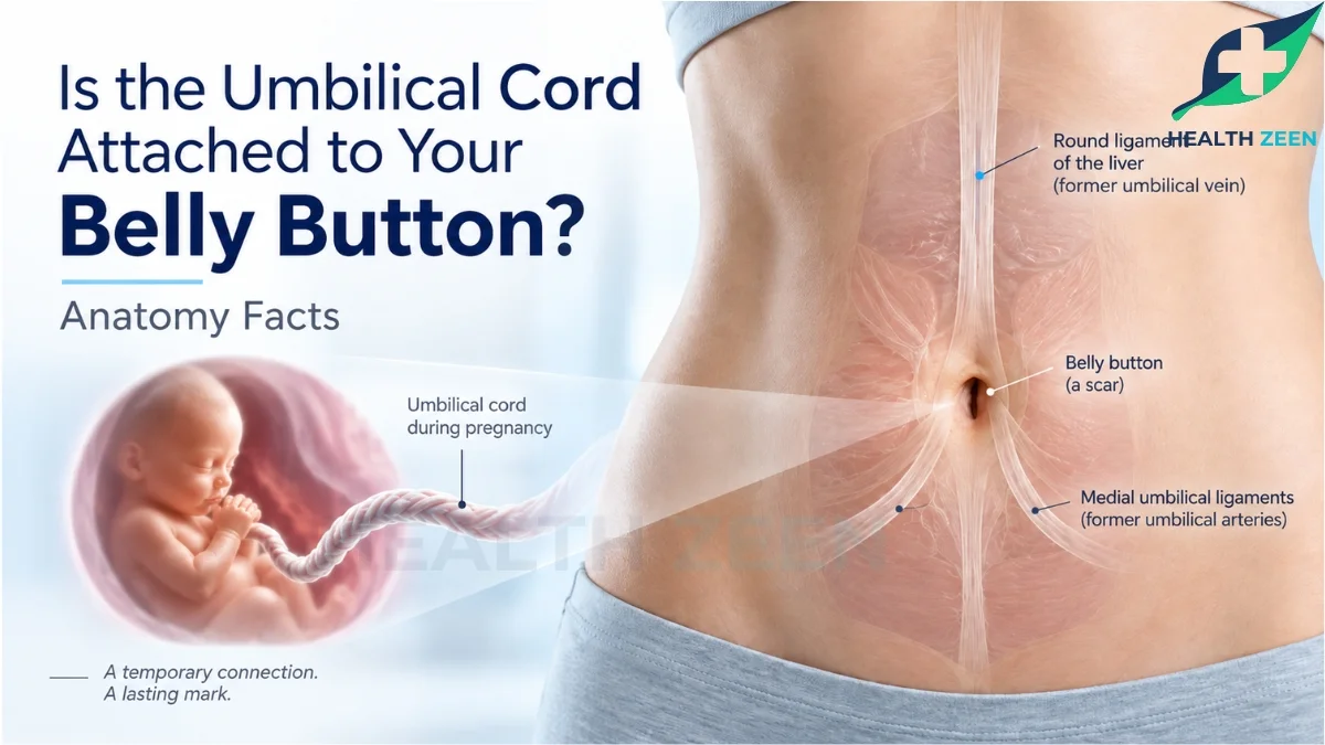
The umbilical cord is no longer attached to your belly button, because your belly button is actually the permanent physical scar left behind after the cord was severed at birth. During your entire developmental period in the womb, the umbilical cord was attached directly to the exact center of your abdomen, serving as the critical biological pipeline between your developing body and your mother’s placenta.
Understanding the anatomical relationship between the belly button and the umbilical cord requires looking backward at human fetal development. The adult umbilicus is not just a random indentation on the stomach; it is a profound biological landmark representing your original source of life support. When the cord is cut immediately after delivery, the external portion dies and falls away, but the complex vascular structures that once existed inside the cord undergo a massive transformation deep within the adult pelvic cavity, shifting from active blood vessels to highly specialized internal ligaments.
The Biological Function of the Umbilical Cord
To understand what the belly button represents, you must understand the clinical function of the umbilical cord before it was detached.
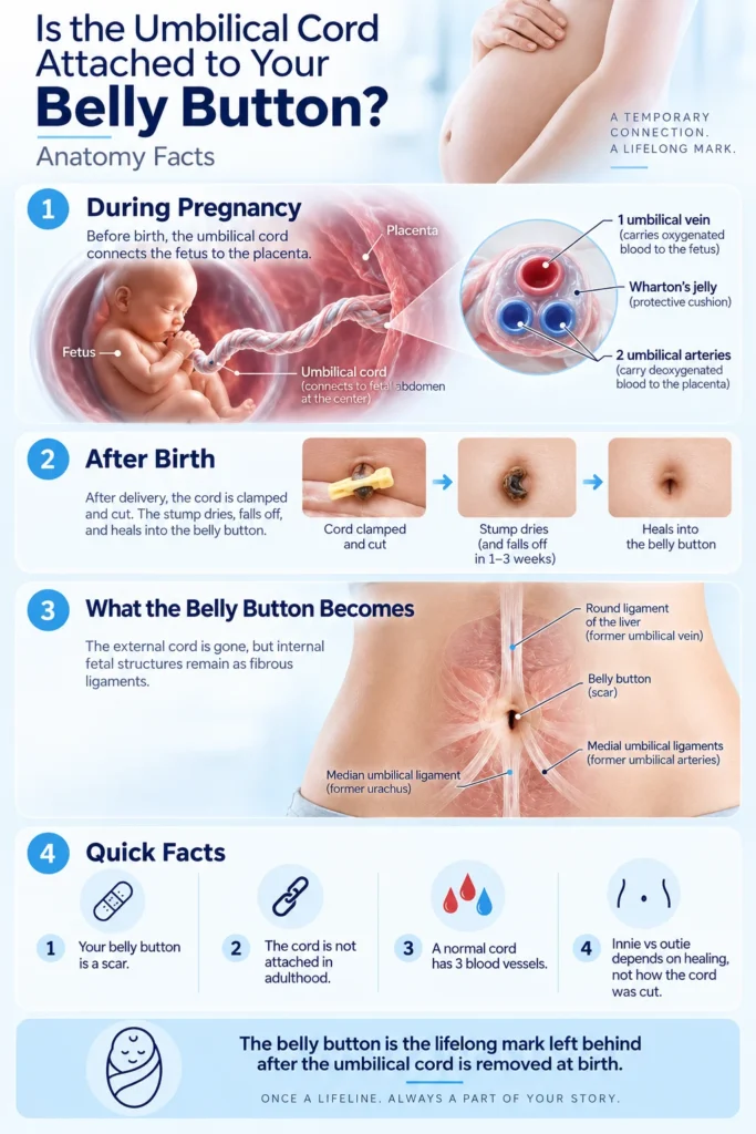
During gestation, the fetus cannot breathe air or digest food. The umbilical cord serves as the sole delivery system for oxygen and nutrients. Clinically, a healthy umbilical cord contains three distinct blood vessels: two umbilical arteries and one large umbilical vein. These vessels are encased in a thick, gelatinous substance called Wharton’s jelly, which protects the lifeline from being compressed or kinked as the fetus moves inside the uterus.
The large umbilical vein is responsible for carrying nutrient-rich, highly oxygenated blood from the placenta directly into the center of the fetal abdomen. Simultaneously, the two umbilical arteries pump deoxygenated blood and cellular waste products away from the fetus, back through the exact same attachment point, and into the mother’s circulatory system for filtration.
The Process of Postpartum Separation
The moment a human is born and takes their first breath, the lungs expand, and the entire circulatory system undergoes a rapid, massive physiological shift. The umbilical cord is no longer biologically necessary.
Physicians clamp the cord to stop the blood flow and painlessly sever it, leaving a small stump attached to the newborn’s abdomen. Over the next one to three weeks, this remaining external tissue naturally dehydrates, oxidizes, and falls off. The circular wound left behind on the anterior abdominal wall gradually heals, closes over with dense fibrous connective tissue, and forms the permanent scar clinically known as the umbilicus, or the belly button.
A common anatomical misconception is that the adult belly button is simply a dead end that connects to nothing. While the external cord is permanently gone, the internal tubes that once connected to it do not disappear; they transform.
Once the umbilical cord is severed, the internal sections of the umbilical vein and arteries suddenly lose their blood flow. Without circulating blood to keep them open, these hollow biological tubes physically collapse. Over several months, the body converts these collapsed blood vessels into dense, fibrous cords known as ligaments. Even in an eighty-year-old adult, these ancient ligaments remain securely attached to the back of the belly button from the inside of the abdominal cavity.
The Round Ligament of the Liver
The massive umbilical vein that once carried oxygenated blood to the fetus collapses and transforms into a thick fibrous band called the round ligament of the liver, or the ligamentum teres hepatis. This strong internal cord runs directly upward from the back of your adult belly button and anchors itself deeply into the tissue of your liver.
The Median and Medial Umbilical Ligaments
The two umbilical arteries that carried waste away from the fetus collapse and transform into the medial umbilical ligaments. These cords run downward from the back of the navel into the deep pelvic cavity. Additionally, a fetal tube called the urachus, which once connected the umbilical cord to the developing urinary bladder, collapses to form the median umbilical ligament, which permanently anchors the adult bladder to the posterior wall of the belly button.
Does the Cord Determine an Innie or an Outie?
Many adults mistakenly believe that the physical appearance of their belly button is determined by how the doctor cut or clamped the umbilical cord at birth. This is entirely a medical myth.
The physician merely cuts the cord a few inches away from the skin. The final shape of your belly button is determined entirely by how your own body heals the remaining fascial tissue. As the abdominal muscles close together during the first few months of life, the scar tissue naturally pulls inward for the vast majority of humans, creating a recessed innie. If the central abdominal fascia does not close tightly, or if a minor umbilical hernia occurs during the healing process, the scar tissue is pushed outward, resulting in an outie.
Frequently Asked Questions
During fetal development, the umbilical cord was directly attached to the center of your abdomen. After birth, the cord is cut and falls off, leaving behind a permanent scar that we call the belly button.
In an adult, the inside of the belly button is connected to several dense internal ligaments. These include the round ligament of the liver running upward, and the median and medial umbilical ligaments running downward toward the bladder.
The vast majority of humans have a belly button because every fetus requires an umbilical cord for survival. However, some adults who have undergone severe abdominal surgeries or specialized hernia repairs may have had their umbilicus surgically removed.
How many veins are in the umbilical cord?
A healthy, normal umbilical cord contains exactly one large vein and two smaller arteries. The vein delivers oxygen and nutrients to the fetus, while the two arteries carry cellular waste and deoxygenated blood back to the placenta.
Yes, strictly speaking from a clinical dermatology perspective, the umbilicus is nothing more than a centralized, permanent scar marking the exact location where your biological lifeline was severed from the abdominal wall.
No, an outie belly button has absolutely nothing to do with how the doctor cut or clamped the umbilical cord. Outies are formed by the natural healing dynamics of your abdominal fascia and the underlying connective tissue.
What happens to the umbilical cord inside the body after birth?
Inside the body, the blood vessels that connected to the umbilical cord immediately collapse after birth. The body permanently converts these collapsed tubes into thick, fibrous ligaments that help anchor the liver and the bladder.
You cannot easily feel these internal ligaments by simply pressing on your stomach, as they are located deep behind the abdominal fascia. However, severe pulling or stretching of these ligaments during pregnancy can cause noticeable localized pain.
The belly button is highly sensitive because the skin forming the scar is exceptionally thin, and it rests directly over a dense network of spinal nerve pathways, specifically the T10 dermatome, which registers sharp sensations when compressed.
In a fully developed adult, the belly button serves no active physiological purpose. However, modern surgeons frequently utilize the umbilicus as a discrete, natural entry point for minimally invasive laparoscopic surgeries to avoid creating visible abdominal scars.
Conclusion
Your belly button is a permanent biological monument to your time in the womb. While the physical umbilical cord is completely detached and discarded shortly after birth, the intricate anatomical connections it forged remain with you for a lifetime. The umbilicus is far more than a simple surface scar; it is the central anchor point for a complex network of internal ligaments that once functioned as your primary arteries and veins. Understanding that the round ligament of the liver and the median umbilical ligament still physically tether your internal organs to the back of your navel demystifies the human anatomy, proving that the legacy of the umbilical cord extends far beyond the delivery room.
Disclaimer
This blog is for informational & educational purposes only, and does not intend to substitute any professional medical advice or consultation. For any health related concerns, please consult with your physician, or call 911.